LociOiling (talk | contribs) (Clarify missing cysteine and methionine.) |
LociOiling (talk | contribs) (2nd attempt.) |
||
| Line 13: | Line 13: | ||
This gallery is based on this [http://www.cryst.bbk.ac.uk/PPS2/course/section7/os_hres.gif hydrogen bonding diagram], from Birkbeck College's online [http://www.cryst.bbk.ac.uk/PPS2/course/ Principles of Protein Structures]. |
This gallery is based on this [http://www.cryst.bbk.ac.uk/PPS2/course/section7/os_hres.gif hydrogen bonding diagram], from Birkbeck College's online [http://www.cryst.bbk.ac.uk/PPS2/course/ Principles of Protein Structures]. |
||
| − | Note that the two sulfur-containing sidechains, [[Cysteine|cysteine]] and [[Methionine|methionine]] aren't included here. The sulfur in methionine doesn't form additional bonds, while the sulfur at the tip of cysteine can form a [[Disulfide Bridge|disulfide bridge]], which is type of [[Covalent bond|covalent bond]]. Covalent bonds are much stronger than hydrogen bonds. |
+ | Note that the two sulfur-containing sidechains, [[Cysteine|cysteine]] and [[Methionine|methionine]], aren't included here. The sulfur in methionine doesn't form additional bonds, while the sulfur at the tip of cysteine can form a [[Disulfide Bridge|disulfide bridge]], which is type of [[Covalent bond|covalent bond]]. Covalent bonds are much stronger than hydrogen bonds. |
<gallery navigation="true" hideaddbutton="true"> |
<gallery navigation="true" hideaddbutton="true"> |
||
Revision as of 03:25, 16 December 2017
The Amino Acid Gallery shows the shapes and chemical composition of each of the 20 amino acids used in Foldit.
Many of the amino acid sidechains contain oxygen and nitrogen atoms that can act as hydrogen bond donors or acceptors.
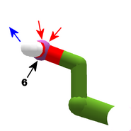
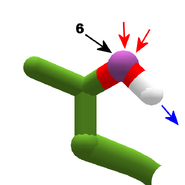
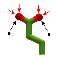
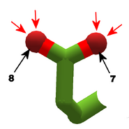
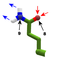
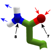
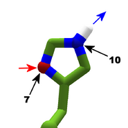
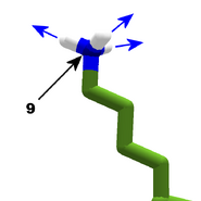
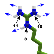
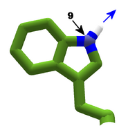
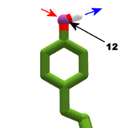
In View Options, the "show bondable atoms" option, combined with "show bonds (sidechain)", highlights the donors with blue spheres, and acceptors with red spheres. Some atoms can act as either a donor or acceptor, which is shown with a purple sphere.
The images in this gallery show the sidechains with bondable atoms. The view used is a combination of "cartoon thin" and "stick plus polar H". The "polar H" option reveals the hydrogen atoms, which normally aren't shown. In this view, hydrogens appear as white "caps" near a blue nitrogen or a red oxygen atom. The hydrogens are of course what make the nearby "heavy" atom (nitrogen or oxygen) a hydrogen bond donor.
Blue arrows show possible hydrogen bond donations (one per hydrogen atom). Red arrows show where hydrogen bonds can be accepted.
Black arrows and numbers show the bondable heavy atom. (In the last segment of a protein section, known as the C terminal, add one to the atom number shown.) The atom numbers can be used when creating bands in a Foldit recipe. The Sidechain Bonding Table summarizes the bondable atoms.
This gallery is based on this hydrogen bonding diagram, from Birkbeck College's online Principles of Protein Structures.
Note that the two sulfur-containing sidechains, cysteine and methionine, aren't included here. The sulfur in methionine doesn't form additional bonds, while the sulfur at the tip of cysteine can form a disulfide bridge, which is type of covalent bond. Covalent bonds are much stronger than hydrogen bonds.
